 Image credit: G. Reddy
Abstract
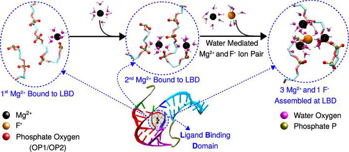
Riboswitches sense various ions in bacteria and activate gene expression to synthesize proteins that help maintain ion homeostasis. The crystal structure of the aptamer domain (AD) of the fluoride riboswitch shows that the F– ion is encapsulated by three Mg2+ ions bound to the ligand-binding domain (LBD) located at the core of the AD. The assembly mechanism of this intricate structure is unknown. To this end, we performed computer simulations using coarse-grained and all-atom RNA models to bridge multiple time scales involved in riboswitch folding and ion binding. We show that F– encapsulation by the Mg2+ ions bound to the riboswitch involves multiple sequential steps. Broadly, two Mg2+ ions initially interact with the phosphate groups of the LBD using water-mediated outer-shell coordination and transition to a direct inner-shell interaction through dehydration to strengthen their interaction with the LBD. We propose that the efficient binding mode of the third Mg2+ and F– is that they form a water-mediated ion pair and bind to the LBD simultaneously to minimize the electrostatic repulsion between three Mg2+ bound to the LBD. The tertiary stacking interactions among the LBD nucleobases alone are insufficient to stabilize the alignment of the phosphate groups to facilitate Mg2+ binding. We show that the stability of the whole assembly is an intricate balance of the interactions among the five phosphate groups, three Mg2+, and the encapsulated F– ion aided by the Mg2+ solvated water. These insights are helpful in the rational design of RNA-based ion sensors and fast-switching logic gates.